


University researchers first to document molecular cancer process
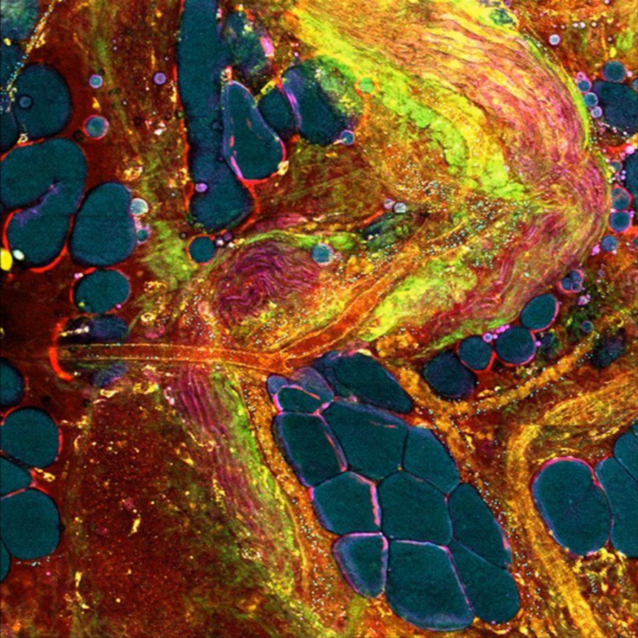
Photo courtesy of Stephen A. Boppart.
A microscopic view of a cell, made possible by University researchers.
February 9, 2017
Accidents happen all the time; however, not all accidents are a bad thing. Researchers at the University who recently stumbled upon new information in cancer research.
The group specifically had a breakthrough in how tumor cells communicate with one another and metastasize throughout the body.
Stephen Boppart, professor of engineering and director of the Center for Optical Molecular Imaging, said the discovery was made possible by a newly developed microscope at the Center for Optical Molecular Imaging.
“We develop optical imaging technology and new microscopes, new ways of using light to look at molecules or cells or tissues and cancer is one of those topics,” Boppart said. “We developed a new type of microscope that allows us to see these little exports.”
The microscope developed by Boppart and his team contains a laser that produces a specific light that enhances visuals not caught by the naked eye. They were experimenting with the microscope on cancer tissue samples one day when they started to notice blue dots within the tissue. After looking into other research, they came to believe that these were tumors that were previously unable to be seen by scientists.
Get The Daily Illini in your inbox!
Historically, scientists and researchers have thought that tumor growths are a product of mutations which initiates them to divide and spread throughout the body. However, they are now starting to understand that there is a different process that involves these tiny “exports.”
As tumor cells start to grow and divide, they produce exports that are spread throughout the body. Exports are bundles of DNA or molecules that transfer to cells, which cause them to change their behavior and metabolism. Essentially, the exports are pre-conditioning the rest of the tissue in the body to be more receptive to the tumor cells.
Haohua Tu, senior research scientist at the Beckman Institute for Advanced Science and Technology, emphasized that the process is microscopic, even on a molecular level.
“The vesicles are very small; they are nanometer sized,” Tu said.
Boppart said the exports can be as small as 30 nanometers. Compared to a cell, which is 10 microns, the exports are about one million times smaller.
The Center for Optical Molecular Imaging has a system that can image the exports in vivo, which means looking at a tissue within a living subject. This allows researchers to capture, in real time, where the exports originated, their destination and effect on the cells.
Sixian You, Ph.D. graduate student in bioengineering, is excited about the potential of the imaging systems.
“In terms of engineering, we want to make the microscope faster with imaging in larger areas,” said You. “We’ve done a lot of increments to the microscope so we can do live imaging with a living rat.”
As this is just the beginning, there is a lot to learn about the fundamental process of cancer before this information will be used for treatment of cancer patients. There are potential targets for therapies. Being able to image these allows for a lot more studies to be conducted.
“These types of discoveries only come about from interdisciplinary research” Boppart said. “It really takes all these people from all these backgrounds to come together to solve these types of problems. It’s important to think how your education relates to other