Research speeds breast cancer biopsies
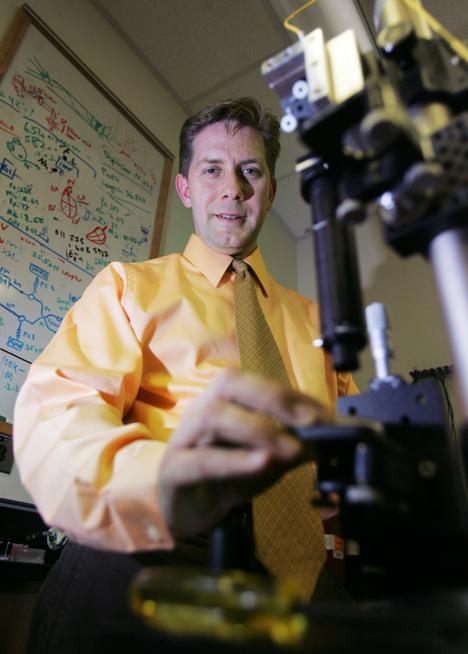
Stephen Boppart, a member of the Beckman Institute Nanoelectronics and Biophotonics Group as well as an associate professor in electrical and computer engineering and bioengineering, is at the forefront of breast cancer-detecting technology. To his right Josh Birnbaum
Oct 5, 2006
Last updated on May 12, 2016 at 05:06 a.m.
Researchers at the University are developing a way to use optical imaging to diagnose and treat breast cancer.
“We want to create technology to do optical biopsies,” said Stephen Boppart, a research physician at Carle and a professor of electrical and computer engineering, bioengineering, and medicine at the University. “This would let us perform a biopsy without actually taking the tissue out.”
The technology that Boppart and colleagues are working with is called optical coherence tomography; it uses near-infrared wavelengths to create images.
“It is the optical equivalent of ultrasound,” Boppart said. “Instead of putting sound waves in, we put in light and track how it is reflected back”
Get The Daily Illini in your inbox!
Boppart does translational research, which aims to translate technological knowledge to real-life clinical applications.
“The National Institutes of Health and the University are interested in translational research because there are lots of good ideas out there, but we need to take our understanding and make it useful to patients,” Boppart said.
Boppart said that while there are a number of applications for this technology, they are focusing their efforts on breast cancer because of its prevalence.
According to 2006 cancer statistics from the American Cancer society, it is estimated that 212,920 women and 1,720 men will be diagnosed with breast cancer this year. Approximately 9,250 of these cases will occur in Illinois alone. By the end of this year, 41,430 people will die of breast cancer, including 1,780 women in Illinois.
The current way to diagnose breast cancer is by performing a needle biopsy in the problematic region as indicated by a mammogram. This procedure requires that a needle be inserted into the breast to remove a small sample of tissue that is then examined under a microscope by a pathologist.
Optical coherence tomography can speed up this process. Light can be delivered through a fiber the size of a human hair to create an image projected in real time, so that the doctor can see it while inserting the needle.
The breast is made of fat cells and stroma, a type of fibrous connective tissue that surrounds fat cells. Tumors are far denser than either of these cell types, so they stand out in the projected image.
Some members of Carle Foundation Hospital are highly enthusiastic about the progress being made.
“We’re very excited about this technology,” said Cathy Emanuel, vice president of business development at Carle. “If it’s developed into a standard treatment, it would be enormously beneficial. It would help surgeons figure out where they need to cut, and give a potentially better outcome.”
With current needle biopsy technology, about 15 percent of cases come back as non-diagnostic, Boppart said. This means that although the mammogram indicates a mass, the tissue comes back as normal. In these cases, surgery is required for a diagnosis.
“We hope to eliminate the non-diagnostic sampling rate,” Boppart said. “That way, we can get a diagnosis without sending the person to surgery.”
When a tumor is removed from a patient, the surgeon also cuts out a portion of normal tissue surrounding the tumor mass to make sure that there are no cancer cells left. The tissue is sent to be microscopically examined by a pathologist.
“The whole process is really time consuming,” Boppart said. “We want to put a portable optical coherence tomography system into the operating room.”
This would allow surgeons to see the tumor margins right away, bypassing the steps of removing tissue and sending it to the pathologist.
“It can save money, time, multiple operations, and even the anxiety of waiting for the patient,” Boppart said.
The technology can be used in the later parts of surgery to identify metastatic spread to the lymph nodes.
The lymph nodes are the body’s natural filter system, so if cancer spreads, it is likely to go to the lymph nodes.
Boppart wants to use optical imaging to check the lymph nodes without having to actually remove them.
“Right now we are working with oncologists, surgeons and pathologists at Carle to see if the optical imaging gives the same results as histology (the microscopic examination of the tissue),” Boppart said.
“Once we determine our images are equivalent to histology, we will know this technology will have a significant impact in medical diagnostics,” he added.


