3D Cow Anatomy App Developed for Veterinary Students
Nov 8, 2015
Last updated on May 5, 2016 at 07:35 p.m.
When she worked at the University, Janet Sinn-Hanlon, medical illustrator in Veterinary Medicine, worked alongside bioengineering professor Gerald Pijanowski.ss The two tried to create animations that would help students visualize animal body parts. They used CAT scans and MRIs to develop an interactive hind leg of a dog on a computer. But the programs they used made the animation results dense, making it difficult to add textural aspects to the animations.
Alan Craig, associate director in Computer-Human Interaction, worked to create augmented reality applications that were published in National Center for Supercomputing Application magazine.ss Opening a special app on a mobile device, viewers could see and interact with a dinosaur and DNA molecules just by looking through the app onto the magazine page.
Upon seeing his work, Sinn-Hanlon and Kerry Helms, coordinator of graphic design in Veterinary Medicine, were inspired to team-up with Craig to bring virtual and augmented reality aspects to the classroom for veterinary students.
Get The Daily Illini in your inbox!
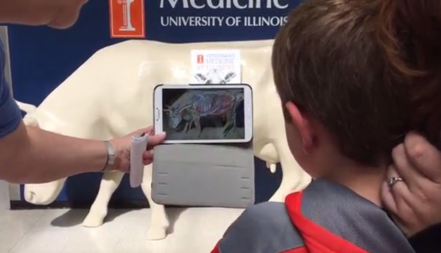
“We’re using computer vision, the tablet, the phone as a magic lens,” Craig said. “We’re looking at the real world through the camera of this device.”
The app takes in information about the position of the magazine page and the position of the viewer to construct an image through the lens of the device. Viewers can move around the virtual image in order to see the various angles of the projection.
In preparation for the College of Veterinary Medicine’s Research Day last Aprilss, the team developed posters that helped showcase the technology. Using the corresponding app, viewers watched a 3-D movie of the anatomy of a dog eyeball and a cow anatomy figure.
The team started to think of ways they could use the technology in an educational way. Pijanowski also had a goal to help students understand the concepts of anatomy in the 3-D sense.
“That’s difficult, especially on a really large animal. You’re used to looking at a textbook and an illustrator might have several views all on one page,” Helms said. “But to really put that all together in your head is kind of challenging. So the exciting part for us is the students can actually look at this and turn it around on their own.”
The app can be used on certain 3-D images to maneuver on a device with respect to the 3-D animation. The new app for the 3-D cow allows students to walk around the image and get a continuous image of the cow’s internal anatomy from any angle.
“There is so much stuffed inside of a body. It’s very compact,” Sinn-Hanlon said. “If you’re going to go on and do surgery, it’s kind of difficult to see where (vessels and nerves) go. It is so helpful to see something in 3-D and rotate things around.”
Veterinary students learning anatomy are already experimenting with some aspects of the technology in classrooms.
In the future, worksheets and booklets would be given to students so they have access to the 3-D anatomy models without having to be in a lab or on a farm. Given the wide variety of animals veterinarians work with, the team is in the process of thinking of different animal anatomies they can re-create to better the learning environment.
“To find where to put the stethoscope on a cow to hear the heart, (students) have to find the elbow, go behind it, count the ribs, and then they have to put it at a certain rib number,” Sinn-Hanlon said. “To have the actual organs behind the large fiberglass cow model, which they use for teaching, was very helpful.”


