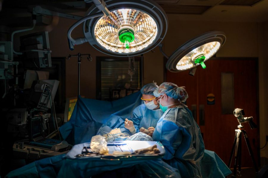
University researchers illuminate cancer tissue in the operating room
Breast surgeon Julie Margenthaler, MD, and clinical fellow Diana Hook (teal mask), MD, perform surgery on March 26, 2018. Margenthaler is co-author on a paper about a camera sensor based on the visual system of a morpho butterfly that can see multi-spectral information.
April 19, 2018
Over the past four years, researchers at the University have developed a camera, inspired by the morpho butterfly, that illuminates cancerous tissue for surgeons to better detect and remove during surgeries.
Viktor Gruev, lead researcher and Engineering professor, said they examined the nanostructures in the morpho butterfly’s eyes to create the camera.
“We want to be able to detect tumor-targeted drugs that are fluorescent during the surgery,” Gruev said.
Gruev said the nanostructures allow the butterflies to see multiple colors, which provides better optical performance.
He added when the majority of physicians perform cancer surgery, they typically use sight and touch to identify where cancerous and healthy tissue are located.
Get The Daily Illini in your inbox!
Missael Garcia, postdoctoral researcher on the study, said they insert a chemical into a patient’s body, which causes cancerous cells to illuminate at a wavelength visible to the camera but invisible to the naked human eye.
“This is one of the few technologies that can actually help during the time they are actually resecting,” he said.
Gruev said surgeons are back in the dark ages while performing surgery, and patients are relying on the surgeon’s experience and condition, along with other factors.
Garcia said a problem is other technologies are too big to bring inside the operating room, so they would like something that is portable, making it more practical to use.
“Once you remove (the tumor), the technology helps to know there is nothing left behind, and most importantly, that they don’t remove healthy tissue that could create nerve damage,” Garcia said.
Garcia added most people who die from a cancerous tissue or tumors do not die from the first one. They die from a second or third tumor that spreads after physicians believe they took out the entire tumor.
“This should definitely help with people not having to come back to the operating room because of a successful first surgery,” Garcia said.
Garcia said one way physicians can view the fluorescent tissue by camera is by having it projected on a screen, where they can turn around to view it in the operating room. Another option is to use virtual reality goggles.
Garcia said their clinical trials have been going successfully when working with animals and humans.
“In two patients, the physician thought they were done, but our technology captured it and then we removed the tissue,” Garcia said.
Gruev said in this phase of clinical trials, they focused on breast cancer, but the imaging and drugs could be used in other types of solid cancers.
Garcia explained they are going into the second phase of testing, which should occur by the end of 2018. They filed for a patent that is expected to take one to two years to be approved, but they first need to wait on the FDA’s approval of particular drugs.
Gruev said the camera is more sensitive than most FDA-approved instruments.
Stephen Boppart, Engineering professor, said in an email it is critical to determine what is normal physiology and what is abnormal pathophysiology in cancer research.
“Technologies and cameras like this enable us new capabilities for both detecting these differences as well as fundamentally understanding the process of carcinogenesis,” Boppard said.
Gruev said compared with other FDA-approved instruments, they have focused on making the technology smaller for the operating room, reducing costs and adding sensitivity and accuracy to the instrument.
“We’re really pushing this technology to a different frontier,” Gruev said. “It is a quite radically different technology.”


