


Illinois researchers’ development of imaging introduces new health applications in study of Alzheimer’s disease
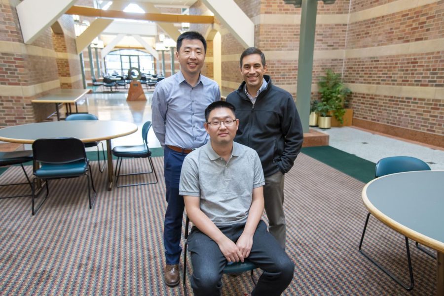
Photo courtesy of the Beckman Institute Office of Communication
Professors Pengfei Song and Daniel Llano pose with graduate student Qi You who aided in Song and Llano’s research. Song and Llano collaborated in developing ultrasound imaging tools that can help study Alzheimer’s disease.
August 4, 2022
Pengfei Song, assistant professor in Engineering, has continually worked on ultrasound imaging technology at the University of Illinois. He described the process through an analogy wherein he and others build hammers, look for the nails and ask themselves, “What is this technology good for?”
Meanwhile, Daniel Llano, neuroscientist and associate professor in LAS, has focused on research projects surrounding different aspects of brain physiology, Llano’s specialty being Alzheimer’s disease.
However, through the Beckman Institute for Advanced Science and Technology, Song and Llano have had the opportunity to collaborate to develop ultrasound imaging tools that can help study Alzheimer’s disease in ways that have not been done previously.
“Beckman houses people from all over campus, so this is an ideal environment for us to explore applications for our technology,” Song said. “I think one thing that this technology is really good at is to see the minute vessels in tissue, and then naturally, we found Dan, who was a neuroscientist who probably at that time needed a tool like this to explore what he’s interested in, so that started the conversation.”
Llano highlighted how essential and beneficial the collaboration with Song has been because optical imaging, the type of imaging that he did prior to working with Song, has limitations. More specifically, optical imaging uses light to image the brain, but since light does not go deep into the brain, Llano was limited to surface-area imaging.
Get The Daily Illini in your inbox!
“I was floored by the images that Pengfei was showing me — just beautiful images of the detailed microvasculature of the whole brain,” Llano said. “I thought this is going to be a great way to be able to do some really interesting neuroscience studies that nobody else is doing.”
Llano contextualized his and Song’s study in the greater history of the study of Alzheimer’s disease, emphasizing that their research involves relatively new ideas. He also noted that the first study of Alzheimer’s disease started over a century ago when Dr. Alois Alzheimer performed an autopsy on a patient and discovered what was happening with the brain’s pathology.
“It took another 80 years before people really understood what was happening in the Alzheimer’s brain, and the field has been mostly focused on an abnormality of something called amyloid,” Llano said. “Amyloid is a protein that in Alzheimer’s patients is present in excessive amounts. It deposits in the brain.”
According to Llano, most of the treatments for Alzheimer’s disease focus on amyloid since many individuals believe that amyloid causes the symptoms associated with the disease. However, Llano noted that treating Alzheimer’s is not as simple as removing amyloid from the brain.
“The problem has been that many of the new treatments that do remove amyloid from the brain don’t seem to be as effective as people had hoped,” Llano said. “I would say over the past 10 years, there’s been a lot more interest looking at other causes and other problems in the Alzheimer’s brain.”
Llano expanded on one important discovery regarding the brains of Alzheimer’s patients, citing that the blood flow is very diminished in specific parts of the brain.
However, Llano acknowledged that no optimal way for studying the brain’s blood flow exists because the problems are located in the capillaries, which are blood vessels in the vascular system that are the size of a human hair.
“The idea that the vasculature is a potential major site of technology and Alzheimer’s — it’s a pretty new idea, but what’s hampered its development has been the lack of a tool to be able to actually look at those blood vessels,” Llano said, again referencing the imaging that has been done before his work with Song. “You can look at them after the organism has died and do a methodology autopsy specimen, but that’s obviously not going to be very useful to really understand what’s going on in the brain.”
Jeffrey Moore, director of Beckman Institute, echoed Llano’s and Song’s emphasis on building upon existing research to develop new imaging tools for the study of Alzheimer’s, stressing how development of new tools and perspectives brings new discoveries.
“If you look at the history of science, imaging or, in general, tools have been able to really be the advancement that’s needed in order to crack a problem,” Moore said. “Anytime any scientist — and go back to Galileo or whoever you want to — has been able to crack a big problem, it’s because they brought a new tool to that problem, and we’re able to see things differently than they had ever been seen before.”
Song described how his and Llano’s focus has been on preclinical trials, which translates to animal experiments, since clinical trials, which translates to human studies, are still limited. In fact, Song and Llano have been studying the brains of mice, and most of their first year of collaboration was spent figuring out how to best obtain images of the brains.
Song clarified the nature of limited clinical trials and the clinical gap as being similar to the chicken-and-egg conundrum.
“Clinically, this is another chicken and egg problem like, ‘Why don’t you study the vessel? Because we don’t have the tool,’” Song said. “And because people don’t have a tool, if you pitch the idea of studying vasculature, people tend to be dismissive about it.”
As Song and Llano worked with the mice, they reinforced a fact that they’ve reiterated regarding imaging — the fact that optical imaging continues to be limited.
“Even on a small-animal models, optical imaging has difficulties of scanning through the entire brain,” Song said.
Song explained that the difficulties go back to how this type of imaging cannot penetrate the brain deeply. Even so, Song mentioned that they were eventually able to explore another path away from optical imaging and instead utilized better resolution MRI and super resolution imaging for the mice.
Ultimately, Song noted that different types of imaging other than ultrasound imaging can involve issues other than the inability to penetrate the brain, such as long scanning times and restrictions on specimen use. However, Song acknowledged that even ultrasound imaging, the imaging they are working toward and developing, has its own limits but that it remains the optimal type of imaging for progress.
He explained that a concept known as phase aberration can mess with and weaken the ultrasound signal but that there is a specific part of the skull that may be key to accessing more of the brain.
“Through the temporal window, there is opportunity to do this in humans clinically,” Song said. “It provides something that is not available with existing imaging modalities.”
Once they were able to obtain appropriate, clear photos of the mouse brains, Song and Llano focused on the aspect of aging before the topic of Alzheimer’s disease. They compared the blood vessels of younger mice and the blood vessels of older mice and discovered that there were major differences in the blood flow of older mice compared to younger mice and how bendy the blood vessels were.
After Song and Llano confirmed their discoveries, they then moved on to the next stage of research, which is where they are now.
“We’re actively working on, as we speak, our early data, (which) are showing very significant but very specific differences in a mouse model of Alzheimer’s disease compared to normal mice,” Llano said. “We’re finding those differences in regions that correspond to the disease regions in human patients with Alzheimer’s disease, so that’s work that’s ongoing.”
Song’s and Llano’s overall work was recently supported to a further degree by a recent two-year grant they received from the National Institutes of Health that will help fund their research. Moore noted that more people are recognizing collaborative grants as very influential and powerful.
“It’s how to get the most bang for the taxpayer buck,” Moore said. “You really want to make sure that you can make the most progress in that period of time, and any way to accelerate that process, like we were speaking about, is going to give you the most progress for the dollar that is being invested in that short period of time.”
Song and Llano both emphasized the support they’ve received from Beckman and how Beckman has played a key role in their research. Specifically, they highlight how Beckman is a unique space where faculty from different disciplines like themselves can collaborate in the same space as opposed to being siloed in different campus areas.
Moore also cited Beckman’s nature as a space of unique, transdisciplinary and collaborative research, expressing his excitement for what Song and Llano have already accomplished.
“The opportunity to bring not only Dan and Pengfei together but their research teams together is really why Beckman exists,” Moore said. “It’s really a pleasure to see the research progress and to see our experiment — the Beckman experiment, the Beckman Institute experiment of interdisciplinary collaboration — playing out in such an important problem.”
Moore even offered his own thoughts on what form the research could take later down the road, emphasizing early diagnosis and a deeper understanding of science as two potential goals that might be accomplished.
“One possible thing that I could imagine as being what I hope that this accomplishes is diagnostic tools that would be able to identify the possibility of Alzheimer’s in someone who otherwise hasn’t been diagnosed yet,” Moore said. “Another outcome that would be helpful would be to discover aspects that are currently not known, that will allow the science to advance to possibly come up with new targets or new directions as to how that intervention might eventually go.”
When considering what the future of their research might look like, Song discussed the development of the next phase of technology that may include the transition from 2D to 3D imaging, the increase in imaging speeds and other improvements that might allow animals to not have to undergo anesthesia during scanning.
Another potential future development that Song emphasized is the further creation of better animal models.
“We’re collaborating with collaborators at Central Michigan University and also working on that effort — trying to basically have a better technology and better animal model,” Song said.
Llano also discussed his hopes for the future of his and Song’s research.
“Once we understand what’s wrong, it’d be really cool if we could try to fix it,” Llano said. “There’s different treatment modalities that can be useful, and then we can use imaging to measure how those animals improve in terms of their microvasculature, and then beyond Alzheimer’s disease, there’s lots of diseases that involve the microvasculature.”
In Llano’s perspective, the study could expand to stroke, traumatic brain injury and even basic science problems unrelated to disease since all of these can be examined through imaging.
“There’s a lot of different directions to go,” Llano said. “There are technical issues that we’re trying to fix as Pengfei mentioned. Being able to image through the skull is something that we’d like to be able to figure out how to do, so we just need enough resources and time, and we get all sorts of things that we could do with this technology.”